Skip to content

Request a Quote

Ready to take the next step? Request a quote and one of our team will be in touch shortly.

Shaping the Future of Endometriosis
Training

Accelerating Diagnosis Through Better Training

Endometriosis remains one of the most underdiagnosed gynecological conditions, with patients often waiting years before receiving answers. The complexity of symptoms and overlap with other conditions contribute to long diagnostic delays, leaving individuals to cope with chronic pain, fertility challenges, and reduced quality of life. While transvaginal ultrasound is a key tool in detection, training standards are inconsistent and access to structured education is limited. Faster, more standardized training is vital to shorten diagnostic delays and ease the burden on patients and healthcare systems.


190 million

worldwide are estimated to be affected by endometriosis, yet on average diagnosis takes 7-10 years


Accelerate Learning with Simulation Training


92%
Improved Understanding
40%
TVS Skill Improvement
150%
Confidence Increase
Picture2b

Real Ultrasound Cases

hexagon red

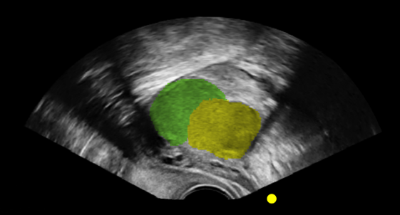
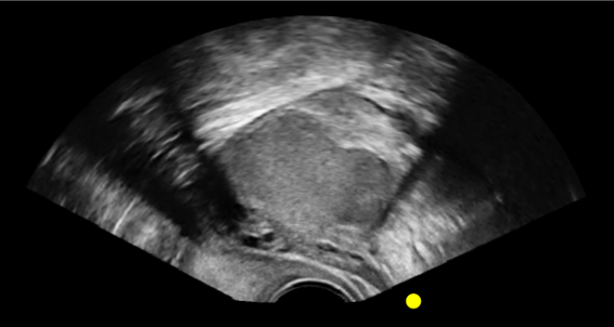
This comprehensive Endometriosis eLearn and simulation module is designed to support clinicians and Allied Health Professionals in developing the skills needed to identify endometriosis during transvaginal ultrasound. Using authentic ultrasound images, the module provides structured, step-by-step training that reflects real-world clinical scenarios and common disease presentations in the pelvis.

The content is organised into four key learning areas:

  • Scanning Technique on a Normal Pelvis – including normal anteverted and retroverted uterus, plus demonstrations of the sliding sign.

  • Endometriosis by Region – 11 interactive cases showcasing varied presentations across pelvic anatomy.

  • Assessment – 5 multiple-choice questions to evaluate scanning skills and knowledge retention.

  • Clinical Case Studies – 12 in-depth cases to consolidate learning, plus supplementary adenomyosis cases* (12 additional studies).


Picture1b

Guided-Learning

hexagon red

Our guided-learning replicates the experience of learning directly from a specialist. Available on each case*, the system provides highlighted ultrasound imagery with clear annotations, allowing learners to gain a clear understanding of what they are looking at, and what to look for in clinical practice – bridging the gap between theory and hands-on practice.

Dynamic highlighting and guided pathways encourage learners to progress at their own pace while reinforcing correct technique. This approach ensures that complex concepts – such as recognising anatomical variations or subtle signs of pathology – are made accessible, consistent, and clinically relevant. With our guided-learning, every scan becomes a repeatable opportunity to build skills under the guidance of an expert.

*Supplementary cases do not include highlighted anatomy


Skip to navigation